What Is an Imaging Test?
What Is an Imaging Test?
Couldn't load pickup availability
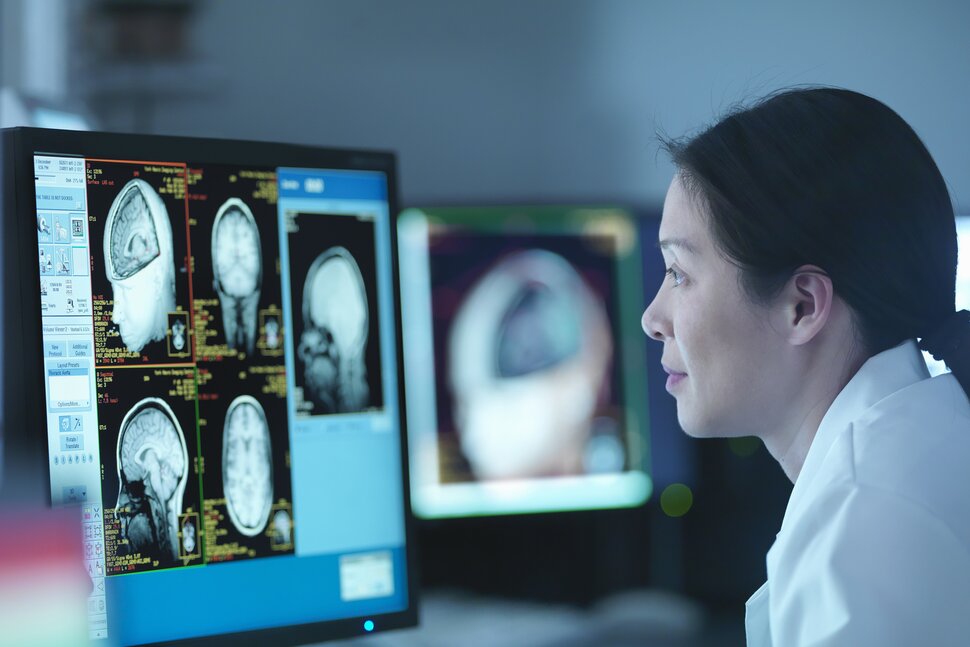
Imaging tests help doctors diagnose and monitor various conditions by visualizing internal structures, organs and tissues. Different types of imaging tests use various technologies, including X-rays, ultrasound, MRIs and radioactive substances (nuclear medicine).
This article is based on reporting that features expert sources.
An imaging test helps health care providers better understand what may be wrong with you so they can make treatment decisions or rule out certain health problems.
There are many different types of imaging tests, so it's easy to get confused about how they work or when they're used.
“Doctors have a range of options when it comes to imaging tests,” says Dr. Evelyn Y. Anthony, professor and chair of the department of radiology at the University of Florida College of Medicine, UF Health Shands in Gainesville, Florida. “In general, we want to use the simplest and least expensive test that will provide the information needed for treatment.”
Here’s more information about the different types of commonly ordered imaging tests.
Common Types of Imaging Tests
If your doctor has ordered a medical imaging exam for you, you might have questions about the type of scan or test you will be having. Technologists use different types of techniques to gather the right images for your radiologist to examine. If you’re getting a scan to see if you have a concussion, for instance, CT would be the modality for your exam. On the other hand, if you are getting a mammogram, X-ray would be the modality in use.
X-rays use a type of electromagnetic energy to get a picture of what’s going on inside the body. This helps health care professionals to see the bones, organs and tissues.
An X-ray is used most often to diagnose conditions such as:
- Fractures
- Pneumonia
- Tumors
- Breast cancer, with the use of a mammogram
Pregnant women are generally advised to avoid X-rays because the quickly developing unborn baby is at greater risk from any radiation harm. Although the amount of radiation exposure from an X-ray is low for adults, it's still good to talk to your provider about any concerns you have about that exposure.
A CT scan (computed tomography scan) blends X-ray and computer technology so health care professionals can see bones, organs, muscles and other parts inside the body. It provides more information than X-rays alone. The scan is taken by moving an X-ray beam around the body in a circular motion.
A CT scan is most commonly used for:
Some CT scans are performed with contrast. Contrast is a special dye taken orally or via injection to show the area being studied more clearly.
Often, health care providers will combine different types of imaging. For instance, they may have an X-ray done to view an ankle injury but then use a CT scan to see the details of a more complex fracture, Anthony explains.
Talk to your health care provider about possibly avoiding a CT scan if you:
- Have kidney problems, as some types of contrast used with a CT scan may lead to kidney failure or
-
Are pregnant
You likely can still have a CT scan with contrast if you’re allergic or sensitive to iodine or shellfish. This is an area of concern because of the ingredients in contrast, but more recent findings have debunked this concern.
Although children can have X-rays, their providers should stay aware of the radiation dose associated with exams like X-ray and CT, says Dr. Vishal Patel, a board-certified radiologist at Stony Brook Medicine in Stony Brook, New York. That’s because too much radiation exposure can raise the risk for certain types of cancer. Your child’s provider can help address the benefits versus risks of imaging tests depending on the reason for having the test.
Magnetic resonance imaging is able to show clear images of everything inside your body, including bones, muscles and organs. Unlike X-rays, MRIs can provide a detailed view of soft tissue areas like the cartilage, ligaments and tendons.
An MRI is done with the use of a very big magnet and radio waves. It's commonly used for:
- Abscesses
- Infections
- Cancer of the brain, spinal cord, breast, prostate, uterus and in some cases, liver
A CT scan, MRI or ultrasound can use two-dimensional (2D) or three-dimensional (3D) imaging. Three-dimensional imaging provides a more detailed view of what’s inside the body.
“3D images help surgeons and radiologists understand the abnormal alignment of bones and how to best plan the surgery. Patients appreciate the simplicity of seeing their imaging test with 3D images,” Patel says.
However, 3D imaging often costs more than regular 2D imaging. Before having an MRI, talk to your healthcare provider if you:
Share

